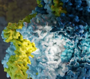
What is mitochondrial dysfunction?
Often referred to as “the powerhouse of the cell,” mitochondria carry out a wide range of functions critical to cellular health. Yet mitochondrial dysfunction can contribute to hundreds of human diseases from cancer to Alzheimer’s, Parkinson’s and mental disorders such as schizophrenia.
Until recently, studies of the ultrastructural details of mitochondria have been limited, especially for statistical studies. Scientists at Scripps Research Institute are combining cryo-electron tomography with a novel computational method to compare their functional differences—leading to new breakthroughs in mitochondria research.
We had the opportunity to discuss these efforts with Danielle Grotjahn, PhD, assistant professor in the Department of Integrative and Computational Biology and Luke Wiseman, PhD, professor in the Department of Molecular Medicine at Scripps Research Institute.
Deconvoluting mitochondrial dysfunction and disease
Dr. Grotjahn heads up the Grotjahn Lab where her team studies the functional and structural interactions that mediate stress-induced modulations to mitochondrial networks.
By using a variety of techniques at the intersection of cellular, molecular, and structural biology, and collaborating cross functionally on mitochondria research with other Scripps Research scientists, including Dr. Wiseman, the lab aims to advance the world’s understanding of physiological and pathogenic mechanisms that contribute to mitochondrial dysfunction and disease.
First, the Grotjahn Lab embraced cryo-electron tomography, cryo-focused ion beam milling and correlative light and electron microscopy to obtain detailed 3D images of mitochondria at nanometer resolution.
“We want to know how all the parts come together,” said Dr. Grotjahn during her interview with Thermo Fisher Scientific. “That’s where cryo-electron tomography really shines because it’s one of the few techniques that allows us to look at not only the details of the individual parts, but also all of them holistically together.”
They also value the use of cryo-electron tomography along with other imaging modalities, “to really get that whole picture across scales,” she said. “I think this will be really useful for other applications in cell biology. Pretty much any cellular process you can imagine is dynamic and has lots of different scales and lots of different parts to it. Being able to image on a cellular and molecular level is really going to transform the field of cell biology.”
No two cells are alike: the case for more statistical measurements
Dr. Grotjahn’s and Dr. Wiseman’s teams recently introduced a novel computational toolkit to help rapidly process these images, turning them into precise measurements that could eventually shed new light on the physiological and pathogenic mechanisms that contribute to mitochondrial dysfunction and disease.
Their ‘surface morphometrics toolkit’ enables the detailed mapping and measurement of the structural elements of individual mitochondria, because no two cells are alike.
“The big question is what can you measure in a tomogram,” said Dr. Wiseman. “There are the things you can see with your eye that strike you very quickly [like] mitochondria cristae for example . . . Next, you have to understand what’s really different about them. Not just, ‘they look different.’ That’s not good enough. You have to be able to say, ‘This curvature is different, the junctions are different, the width of the cristae are different.’”
According to Dr. Wiseman, there were many cases where they’d have to manually draw or model what the different membranes looked like to get these types of measurements.
“Danielle found solutions around some of these problems, where now we’re getting discrete measurements,” he said. “We’re getting those junction curvatures; we’re getting cristae widths.”
Combining cryo-electron tomography with this computational method is proving successful for these groups studying the structural differences between mitochondria in cells that are functioning properly to those within cells under stress.
“The key thing for us to really apply this sort of technology was to be able to develop higher throughput strategies, where we can have multiple different images,” said Dr. Wiseman. “We have to be able to have enough images to we can get statistical significance between differences.”
Disrupting mitochondrial diseases
Most recently, the team used this combination to pinpoint the structural changes that occur when mitochondria are subjected to cellular stresses induced by diseases, toxins, infections, and pharmaceutical drugs.
“What we’re trying to do is figure out how our bodies are naturally protecting themselves from different types of pathological insults, things that are associated with different types of diseases,” said Dr. Wiseman. “If we understand how those pathways work, we can find ways to actually hijack them. And by hijacking them, we can develop new therapeutics to intervene in disease.”
Dr. Grotjahn is very excited about the future of cryo-electron tomography, and it shows.
“What’s really exciting about Scripps is that we are equipped with everything that we need to dive into cells and be able to visualize them in a completely new way and with extreme detail. The infrastructure we have here really allows us to do this at a throughput that enables us to make important biological discoveries more quickly and more streamlined,” she said.
Read Dr. Grotjahn’s and Dr. Wiseman’s recent article published in the Journal of Cell Biology >>
//
Leah Lavery is a Sr. Manager, Life Sciences Market Development at Thermo Fisher Scientific.


Leave a Reply