Search Thermo Fisher Scientific
Disclaimer

Clicking the images or links will redirect you to a website hosted by BenchSci that provides third-party scientific content. Neither the content nor the BenchSci technology and processes for selection have been evaluated by us; we are providing them as-is and without warranty of any kind, including for use or application of the Thermo Fisher Scientific products presented.
Invitrogen
HLA-DR Monoclonal Antibody (Tü36), Qdot™ 800
{{$productOrderCtrl.translations['antibody.pdp.commerceCard.promotion.promotions']}}
{{$productOrderCtrl.translations['antibody.pdp.commerceCard.promotion.viewpromo']}}
{{$productOrderCtrl.translations['antibody.pdp.commerceCard.promotion.promocode']}}: {{promo.promoCode}} {{promo.promoTitle}} {{promo.promoDescription}}. {{$productOrderCtrl.translations['antibody.pdp.commerceCard.promotion.learnmore']}}
")
FIGURE: 1 / 1
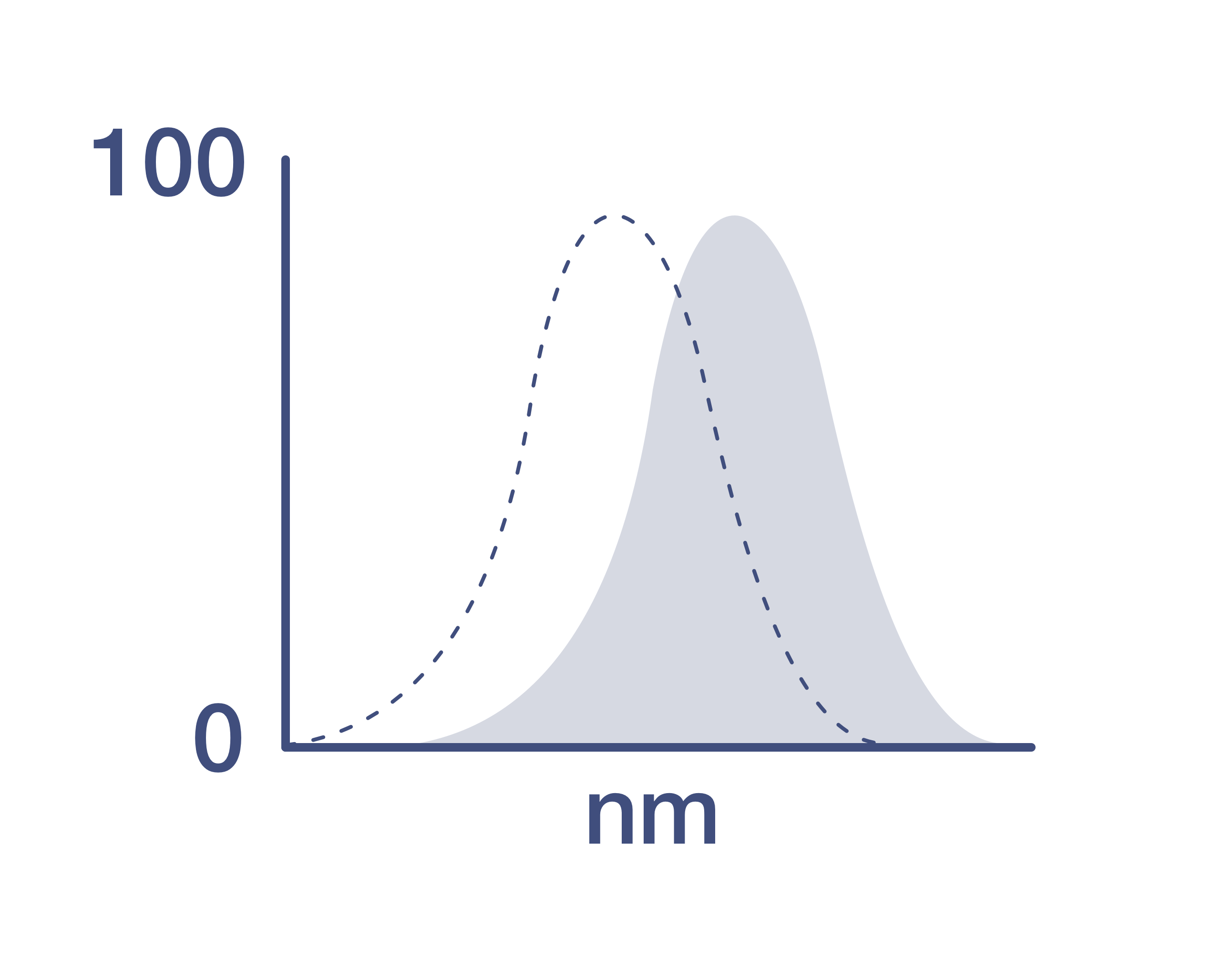
HLA-DR Antibody (Q10063) in Flow
Log fluorescence intensity profiles of human PBLs gated on lymphocytes and analyzed on a BD LSR II flow cytometer (BD Biosciences, San Jose, CA) using 405 nm excitation and the specified emission filters. The data files were analyzed using FlowJo™ software (Treestar, Inc., www.flowjo.com). The black line represents cells stained with anti-human HLA-DR antibody conjugate and the gray line represents unstained cells. Note: Flow cytometric data shown may not necessarily have been generated using the enclosed lot of reagent. For this reason, a... View More

 in Flow")
Product Details
Q10063
Product Specifications
Host/Isotype
Mouse
/ IgG2b
Class
Monoclonal
Type
Antibody
Clone
Tü36
Immunogen
Human HLA-DR
Conjugate
Excitation/Emission Max
300/792 nm
View spectra
Form
Liquid
Storage conditions
4°C
Shipping conditions
Wet ice
RRID
AB_10401405
Product Specific Information
Qdot™ Antibody (Ab) conjugates possess a bright fluorescence emission that makes them well suited for the detection of low-abundance extracellular proteins. Approximately the same size as R-phycoerythrin (R-PE) and compatible with existing organic fluorophore conjugates, Qdot™ Ab conjugates can be excited with any wavelength below their emission maximum, but are best excited by UV or violet light. The narrow, symmetric emission profiles of Qdot Ab conjugates allow for minimal compensation when using a single excitation source, and the very long stoke shifts enable better, more efficient multicolor assays using the 405 nm violet laser. Available in multiple colors for use in flow cytometry, these advantages make Qdot Ab conjugates powerful tools for antibody labeling and staining. Staining: Stain cells in any standard staining buffer, such as phosphate buffered saline (PBS) with 1% bovine serum albumin (BSA). Use 1 L of Qdot antibody conjugate per 1 × 10^6 cells in a 100 µL staining volume (20 nM final concentration of Qdot™ nanocrystals). Qdot™ nanocrystal conjugates may be mixed with other antibodies, but use the diluted conjugates on the day of dilution. Qdot nanocrystal conjugates can be used for surface staining applications with most conventional sample preparation reagents, such as Cal-Lyse™ and FIX & PERM™ reagents, with minimal affect on fluorescence. We have observed that some batches of BD FACS™ Lysing Solution have been found to interfere with Qdot™ nanocrystal fluorescence. Each lot has been tested by flow cytometry using human peripheral blood leukocytes. This testing was performed using 1 L of antibody per 1 × 10⁶ cells in a 100 L staining volume (20 nM final concentration of Qdot nanocrystals). Qdot nanocrystal concentration is assigned based on optical density. The isotype control for this antibody is mouse IgG2a, Cat. No. Q10014.
Instrument setup: Qdot Ab conjugates are excited optimally with UV or 405 nm light, although excitation can be obtained with any wavelength below the emission maximum of a given Qdot nanocrystal, such as with a 488-nm laser. Qdot Ab conjugates can be used on cytometers that do not have UV or violet excitation sources as long as they have appropriate emission filters. Make sure the cytometer has an appropriate emission filter for the Qdot Ab conjugate being used; alternate filters can be used as long as they capture the emission maximum, but filter width impacts spectral overlap corrections. And be sure to check for Qdot Ab conjugate emission in any channel that can capture nanocrystal emission off of other lasers on the cytometer. For Cat. No. Q10063: peak excitation 405 (488) nm/peak emission 800 nm; recommended filter 780/60 nm.
Store reagents at 2-8°C in the dark. Do not freeze. Because Qdot nanocrystals are conjugated to biological materials, some loss of activity may be observed with prolonged storage. When stored as instructed, expires six months from date of receipt unless otherwise indicated on product label. Qdot Ab conjugates are photostable, and do not need to be protected from light. However, if using Qdot Ab conjugates in combination with conventional fluorochrome conjugated antibodies, minimize light exposure during handling, incubation with cells, and prior to analysis. The Qdot Ab conjugates contain cadmium and selenium in an inorganic crystalline form. Dispose of the material in compliance with all applicable local, state, and federal regulations for disposal of these classes of material. For more information on the composition of these materials, consult the Safety Data Sheets (SDSs).
Target Information
HLA-DR, like other MHC class II molecules, is a transmembrane glycoprotein composed of a 36 kDa alpha chain (DRA) and 27 kDa beta chain (DRB). The alpha chain gene contains 5 exons. Exon 1 encodes the leader peptide, exons 2 and 3 encode the two extracellular domains, and exon 4 encodes the transmembrane domain and the cytoplasmic tail. DRA does not have polymorphisms in the peptide binding part and acts as the sole alpha chain for DRB1, DRB3, DRB4 and DRB5. Within the DR molecule the beta chain contains all the polymorphisms specifying the peptide binding specificities. Hundreds of DRB1 alleles have been described and typing for these polymorphisms is routinely done for bone marrow and kidney transplantation. HLA-DR is expressed primarily on antigen presenting cells such as B lymphocytes, monocytes, macrophages, thymic epithelial cells and activated T lymphocytes. Three loci, DR, DQ and DP, encode the major expressed products of the human class II region. The human MHC class II molecules bind intracellularly processed peptides, present them to T-helper cells, and have a critical role in the initiation of the immune response.
HLA and MHC antibodies play a significant role in Immunopeptidomics, facilitating the identification and characterization of neoantigens through high-performance liquid chromatography coupled to tandem Mass Spectrometry.
For Research Use Only. Not for use in diagnostic procedures. Not for resale without express authorization.
How to use the Panel Builder
Watch the video to learn how to use the Invitrogen Flow Cytometry Panel Builder to build your next flow cytometry panel in 5 easy steps.
References (0)
Have you cited this product in a publication?
Let us know so we can reference it here.

Performance Guarantee
If an Invitrogen™ antibody doesn't perform as described on our website or datasheet,we'll replace the product at no cost to you, or provide you with a credit for a future purchase.*
Learn more
We're here to help
Get expert recommendations for common problems or connect directly with an on staff expert for technical assistance related to applications, equipment and general product use.
Contact tech support